WESTERN BLOT
THE WESTERN BLOT

For western blotting proteins are first fractionated by gel electrophoresis using polyacrylamide gels. Usually, denaturing conditions are used, however, non-denaturing conditions can be used during electrophoresis. A very powerful alternative is the use of two dimensional (2-D) electrophoresis that uses the combination of non-denaturing and denaturing conditions. This allows a greater ability to separate similar sized proteins by differences in their intrinsic ionic charge. This is a good method for investigating post-translational modification such as phosphorylation.
The fractionated proteins are then electrophoretically transferred out of the gel to a solid support matrix. Solid supports are sheets of charged polymer membranes that bind proteins through electrostatic and hydrophobic interactions. Once bound the membrane bound proteins can be probed by a variety of methods but most commonly using immunological reactions.
The membrane is incubated with an antibody, called the primary antibody, that binds the target protein. Bound antibodies that remain after washing are then detected with a secondary antibody. This antibody usually is conjugated to a molecule that generates a detectable signal. This molecule is usually an enzyme, either alkaline phosphatase (AP) or horse radish peroxidase (HRP), each having a companion chemical substrate. The product(s) of the enzyme-substrate reaction generate a detectable signal. The signals vary from colored dye precipitates to the generation of light. Secondary antibodies can also be conjugated to molecules such as biotin and fluorophores and detection using streptavidin-HRP or fluorescence detector, respectively.
The Western blot is an assay intended to detect a single protein in a mixture of proteins, similar to the ELISA. Also similar to the ELISA, it requires an antibody specific to the protein that is intended to be detected. However, the Western blot is not performed in a well plate, and involves a separation by molecular weight.
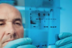
The Western blot involves three steps. First, proteins are separated by size via gel electrophoresis. The gel electrophoresis bands are then transferred to a membrane or solid support. The membrane then is incubated with antibodies specific to the target protein, which are linked to an imaging molecule (either via fluorescence or a colorimetric enzymatic tag). Proteins that have not bound to the antibodies are washed off, and the film is developed to determine the amount and size of proteins that bound with the antibodies.
WES
In addition to the standard Western blot described above, Altogen Labs performs Western blots using ProteinSimple’s Wes Simple Western blotting system. This highly sensitive automated Western blotting system can run 25 samples simultaneously, utilizing a novel capillary system. It can detect protein at the nanogram level in nanoliter-volume samples, decreasing the sample size required for quality control. In addition, its three-hour run time allows us to have same-day verification of protein expression. Use of the Wes allows Protein Expression Services to provide exceptional quality control for our protein expression experiments.
FOR MORE INFORMATION, SEE:
- Request quote for:
- Protein Expression Services
- Protein Expression Applications
- Assay Development
- ELISA
- Western Blot
- Stability Analysis
Laboratory Services │ Science Of Protein Expression │ Protein Expression Applications │ Assay Developmen │ ELISA │ Western Blot │ Stability Analysis │ Available Services